

A Case of the Week
Case 129
An eighty two-year-old female was admitted for respiratory rehabilitation since she had chronic obstructive pulmonary disease and asthma. One week later, she was found in a state of face down on the table in front of the shop at the first floor of our hospital. She could open her eyes and understand what is questioned but her response was not speedy as usual, quite dull. Right upper hemiparesis (upper extremity 1/5, lower extremity 5/5) was noted.
Brain MRI was performed immediately that revealed lesions with high signal intensity at left brain on diffusion weighted imaging (Fig. 1), suspicious of embolism or thromboembolism. She did not appeal to us about visual field deficit but she could not tell the name of the color of things which she watched.
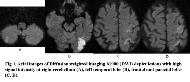
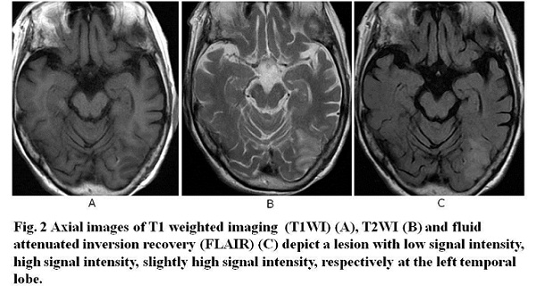
What lobe was damaged in acute infarction on MRI with DWI, T1WI, T2WI and FLAIR (Figs.1B&2)?
2018.11.14
COPYRIGHT © SEICHOKAI YUJINKAI. ALL RIGHTS RESERVED.