

A Case of the Week
Case 201
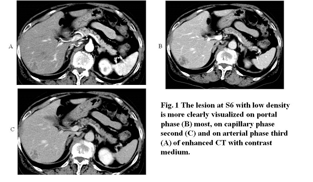
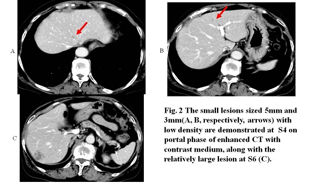
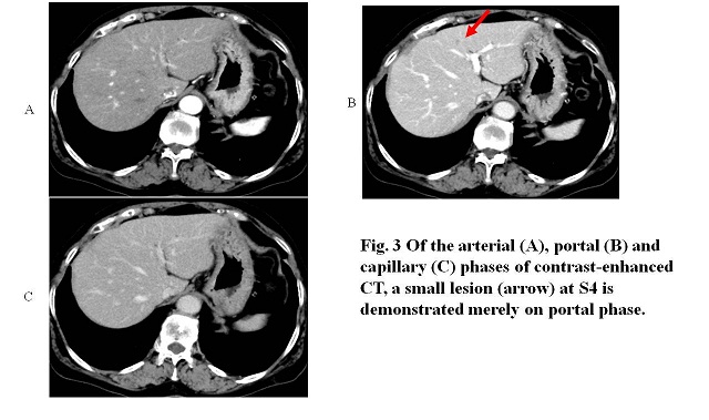
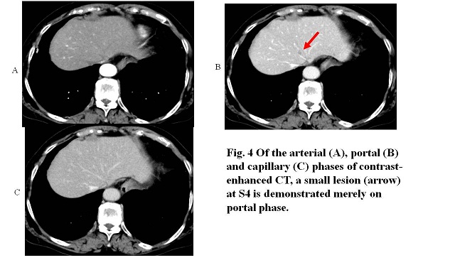
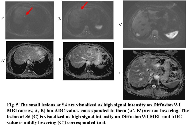
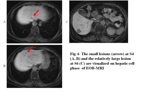
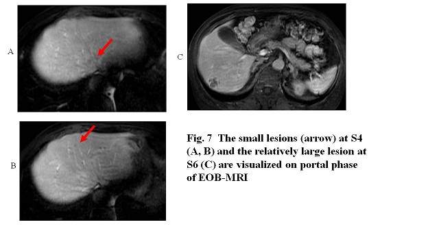
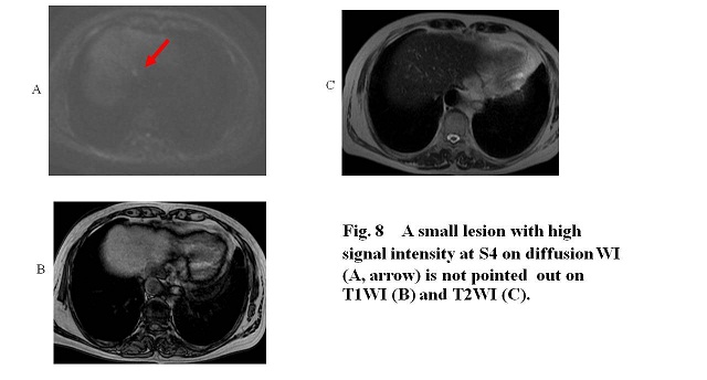
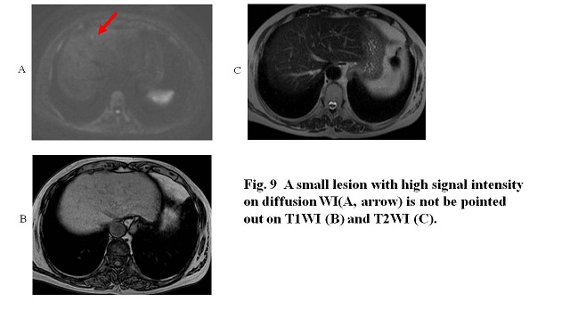
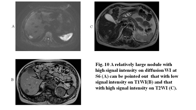
A seventy seven-year-old female periodically (once per two months) presented in our hospital for follow-up after endoscopic intraperitoneal tumor resection for sigmoid cancer three years before. After excision, she was given anticancer drugs. However, distant lung metastasis was found last year and then, she received re-surgical resection for metastatic lung tumor. Laboratory test revealed CEA 3.2 mg/dL and CA19-9, 10 U/mL. She received periodic contrast-enhanced abdominal CT (Figs 1- 4) and thereafter, Gd-enhanced MRI (Figs 5 – 10) to check local recurrence or distant metastasis.
What is the most appropriate imaging on CT to demonstrate the minute liver metastasis ?
What is the most appropriate sequence MRI to demonstrate the minute liver metastasis ?
2020.8.19
COPYRIGHT © SEICHOKAI YUJINKAI. ALL RIGHTS RESERVED.