

A Case of the Week
Case 3
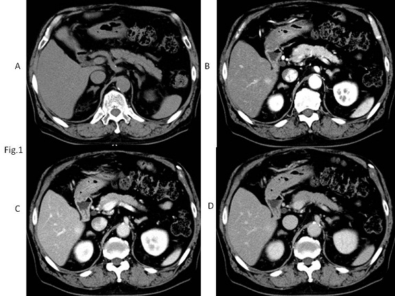
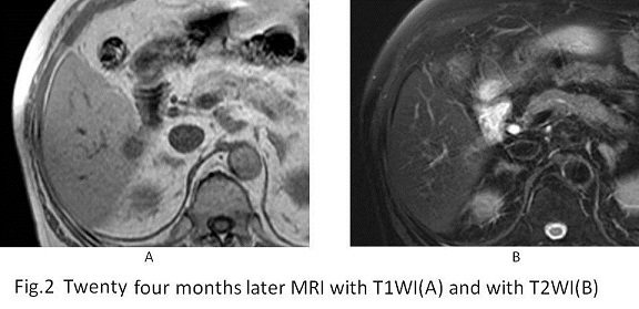
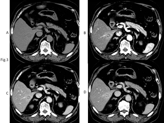
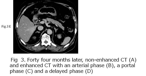
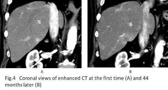
Ten years ago, a seventy four-year-old male had received hepatic sub-segmentectomy for hepatocellular carcinoma. Thereafter, the patient came to our hospital for the follow-up study every year. Five years later, enhanced CT depicted a new lesion with hypervascularity at S6. The lesion gradually grew and had the mixed component of hyper- and hypo-vascularity. Laboratory test revealed no abdominal findings: ALP of 207 U/l, AFP of 3.0ng/ml, PIVKA of 20mAU/ml, CEA of 3.8 mg/ml, CA19-9 of 8U/ml, HCV antibody (-) HBs antigen (-).

What is the diagnosis ?
2016.04.20
COPYRIGHT © SEICHOKAI YUJINKAI. ALL RIGHTS RESERVED.