

A Case of the Week
Case 31
An eighty two-year-old male has routinely obtained rehabilitation in our hospital since he was diagnosed lumbar spinal canal stenosis two years ago. At that time MRI showed osteoporosis, and lumbar disk hernia of L3/4, L5/S1 and thickness of yellow ligament, causing compression of dural sac and equine caudate nerves. In these days, he was getting disable to swallow food smoothly. He was 166 cm tall, 58 kg weight and 21.1 BMI. Laryngeal scope revealed the protrusion of the mucosal surface from the posterior wall of the hypopharynx at the level of the epiglottis. Laboratory tests revealed no abnormal findings. He received neck CT for further investigation.
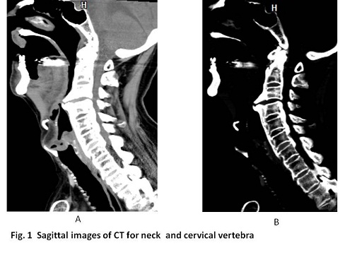
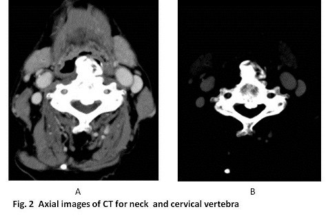
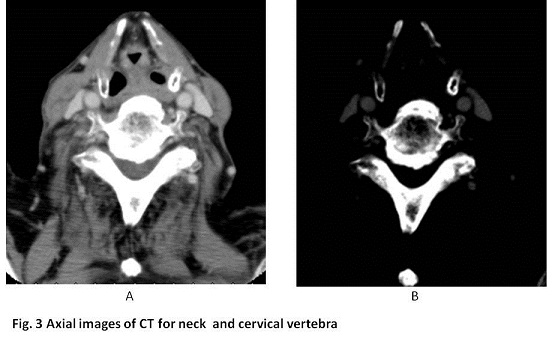
What are the appropriate imaging findings on neck CT ?
2016.11.9
COPYRIGHT © SEICHOKAI YUJINKAI. ALL RIGHTS RESERVED.