

A Case of the Week
Case 44
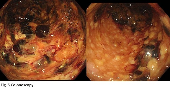
Approximately one year ago, a sixty six-year-old female underwent surgical removal of the right gingival cancer and right cervical lymphadenectomy in our hospital. Four months later, she received left cervical lymphadenectomy for metastatic lymphnodes swelling. Soon after that, the recurrent tumor appeared at the right neck and its volume increased. She was given chemotherapy three times each other month. During the third chemotherapy, intermittent diarrhea of 3 to 10 times per day occurred and continued for several days. Then, third-line chemotherapy was transiently stopped. Colon endoscopy was tried but limited only for rectal examination because of anticipating perforation. Because the tumor was growing aggressively, chemotherapy had to resume. Diarrhea and bloody enema continued. One day, massive rectal bleeding occurred. Laboratory test revealed red blood cells 1430000/ mm3, white blood cells 14340/mm3, neutrophils 91.6 %, hemoglobin 4.3 g/dl, and CRP 0.45 mg/dL.
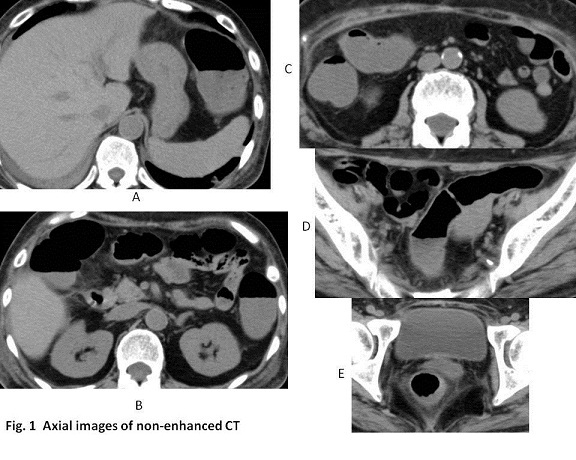
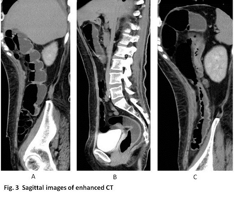
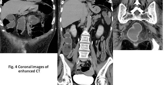
Non-enhanced CT was conducted (Fig. 1) and a few hours later enhanced CT (Figs 2, 3 and 4) was added to check bleeding site.

What is the imaging diagnosis based on the clinical progress ?
2017.3.1
COPYRIGHT © SEICHOKAI YUJINKAI. ALL RIGHTS RESERVED.