

A Case of the Week
Case 55
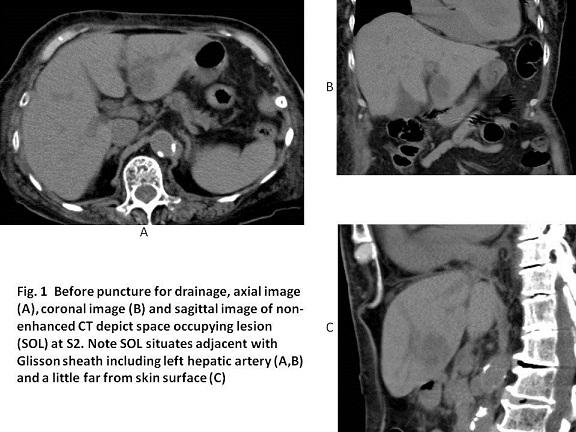
A ninety-year-old female lived alone in her house. She regularly got a public health service. A helper woman measured her body temperature at 6 PM which revealed 38。C and then, the helper woman called her daughter living in Kyoto. Her daughter arrived at her house at 11 PM and measured her mother temperature again which revealed the body temperature rose to 39。C. Then, she called an ambulance car and her mother was transported to our hospital. Laboratory test revealed CRP 26.59 mg/dL, White blood cells 15470/mm3, Neutrophils 90.2%, Creatinin 1.88 mg/dL, platelets 59000/mm3 and procalcitonin more than 10 ng/mL (< 0.5), implying sepsis. Antibiotics and γ-globulin started to infuse. As abdominal distension was found, non-enhanced CT was conducted to make sure what happened. Non-enhanced CT showed low density mass sized 3.2 cm in diameter with unclear margin in the liver (Fig. 1) and echo showed low echoic mass with mosaic pattern, indicating liver abscess.
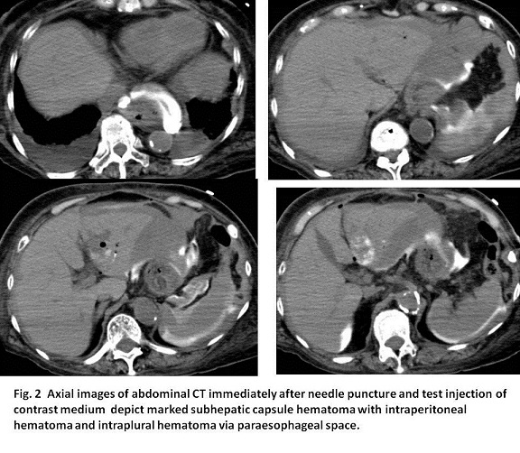
The following day, drainage for liver abscess was attempted under echo and X ray fluoroscoic guidance. Successful puncture was achieved and abscess fluid of 4 ml was taken out. However, drainage tube could not be inserted despite the repeated puncture and at last, blood was coming out via the puncture needle. Test injection via the puncture needle showed contrast medium flowing-out to intraperitoneal space. The blood pressure was getting decreased. Non-enhanced CT showed extrahepatic hematoma (Fig. 2).
【Angiography】
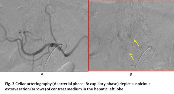
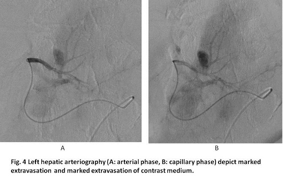
Approximately an hour after the non-enhanced CT, celiac arteriography was conducted using a 3F catheter (Rosch C2). It showed suspicious extravasation of contrast medium in the hepatic left lobe (Fig. 3). Then, a microcatheter using a microguidewire was advanced to the left hepatic artery. Selective left hepatic arteriography showed marked extravasation of contrast medium (Fig. 4).
What is the appropriate management for bleeding from hepatic artery injured by needle puncture for liver abscess ?
2017.5.24