

Clinical diagnosis
Case 62
【Treatment】
Day 1, After paranasal CT was conducted, he was given antibiotics (ceftriaxon) based on imaging diagnosis of paranasal sinusitis.
Day 2, After MRI was conducted, he was transported to university hospital for draining subdural empyema and fluid in the frontal sinuses.
【Discussion】
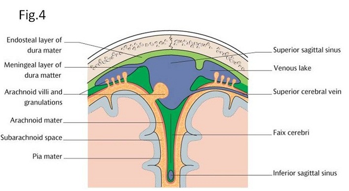
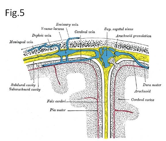
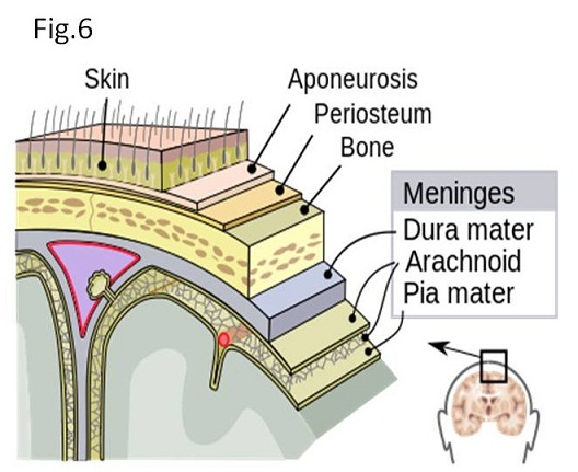
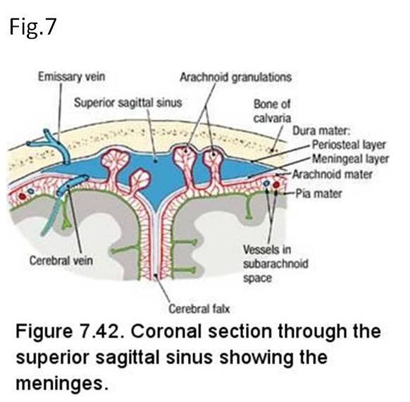
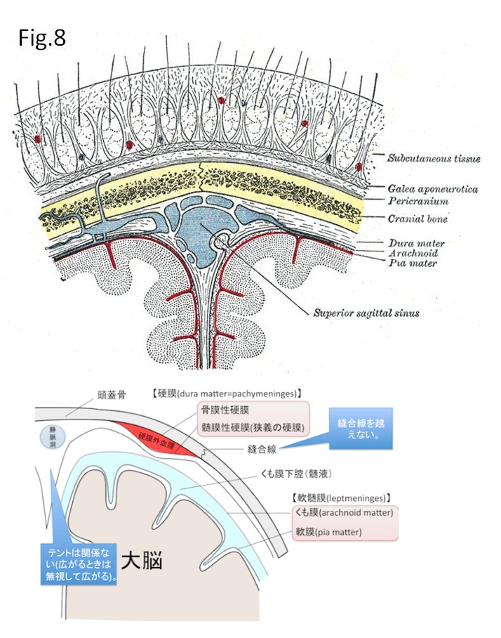
Dura matter has two layers: periosteal layer and meningeal layer (1) (Fig. 4). The two layers look fusion and run together through most of the skull (2 -6). There is a gap between them where they separate, called a dural venous sinus (Figs. 4, 5, 6, 7). For example, superior sagittal sinus situated between them. Dural sinuses drain blood and cerebrospinal fluid from the arachnoid space via bridging vein and flow into the internal jugular vein (Figs. 4, 5, 6, 7) (1). Subdural space exists underneath the meningeal layer of the dura mater, meaning the gap between meningeal layer and arachnoid membrane. Cerebellar tent separates the cerebral occipital lobe from the cerebellum and brainstem, and cerebral falx that is located in the longitudinal fissure, separates the two hemispheres (1). Cerebellar tent and cerebral falx are formed where the periosteal layer and the bilateral separated meningeal layers merge together such as sheet-like protrusions into the cranial cavity (Figs. 4, 5) (1).
Meanwhile, extradural space is a gap between the periosteal layer and skull bone where meningeal arteries branched from the middle meningeal artery exist. It is known that the traumatic skull fracture damages these arteries, causing an extradural hematoma. Extra-dural hematoma occurs associated with skull fracture with the incidence of more than 90 % (7, 8). Extradural space is divided by the suture of cranial bone, while subdural space expands whole brain crossing beyond the suture of cranial bone (2 -6). Then when the lesion, irrespective of hematoma or empyema, crosses beyond the skull fissure, it indicates the subdural lesion.
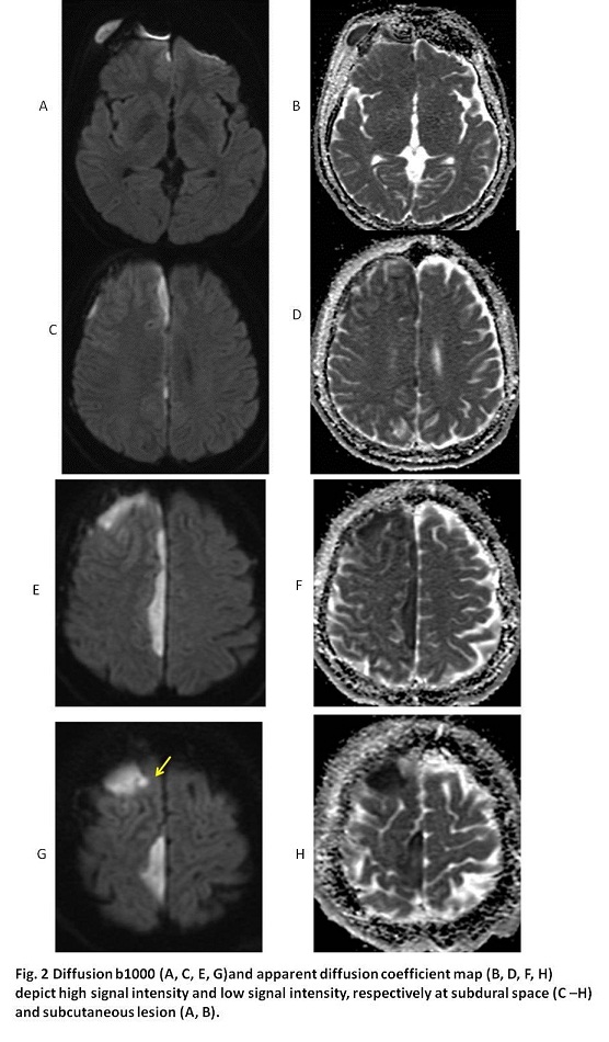
In our case, a longitudinal lesion expands crossing beyond the coronal fissure of skull, implying subdural empyema (Figs. 2C, 2E, 2G). Meanwhile, it seems to be difficult to differentiate whether the left frontal lesion of our case is subdural or extradural because the lesion is localized with the configuration of hemi-convex (Fig. 2G). However, there is a small daughter lesion adjacent to the left hemi-convex lesion (Fig. 2G). It indicates the small brain abscess occurred by intra-parenchymal penetration. Then, the hemi-convex lesion can be interpreted to exist in the subdural space.
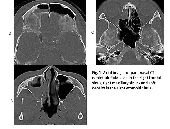
Subdural empyema occurs predominantly in males (approximately 80%) although its reason is unknown (9-11). The occurrence age of two-thirds of patients is 10 to 40 years (9-11). Infection invades to the subdural space directly through the paranasal sinusitis especially through the thin posterior wall of frontal sinusitis or through thrombophlebitis in the venous sinuses. Infection to subdural space can rarely spread from distant foci such as pulmonary abscess or liver abscess. Our case was a thirty nine-year-old male whose symptoms were smelly nasal fluids and fever. Non-enhanced CT showed paranasal sinusitis including frontal sinusitis with thin posterior wall (Fig.1A) which spreads directly subdural space and partially brain abscess.
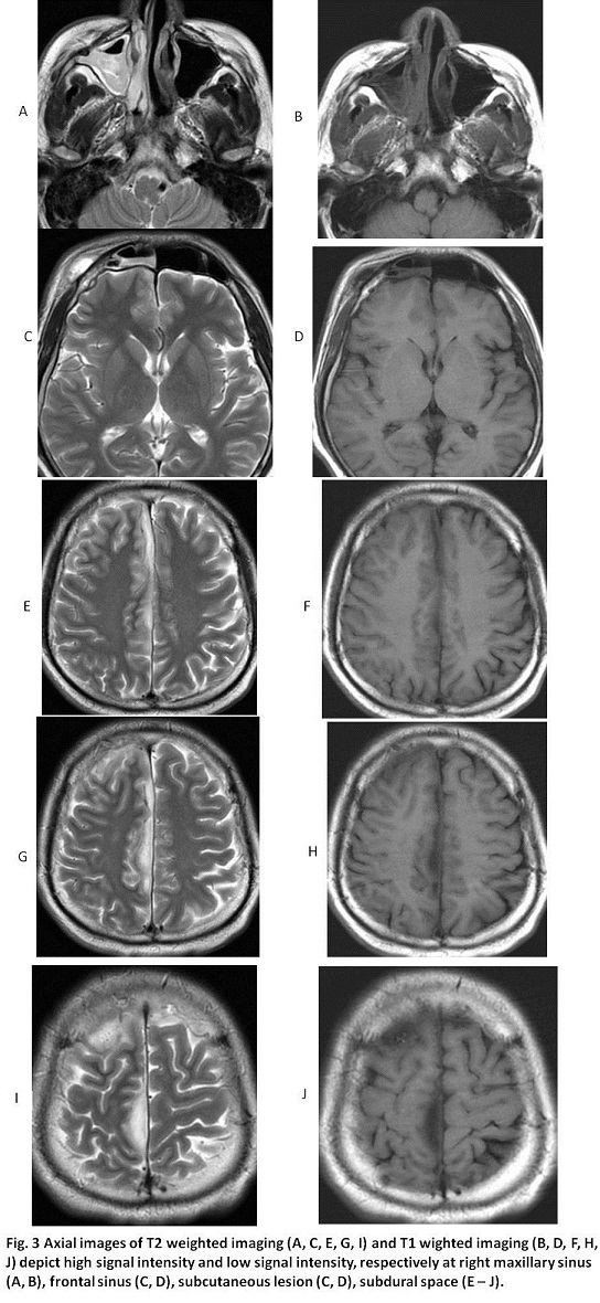
Further, frontal sinusitis spreads directly to subperiosteal lesion of frontal bone (Figs. 6, 8), inducing subperiosteal abscess with the frontal head swelling named Pott puffy tumor (4, -6). In our case, the right frontal eye lid swelling was found and MRI with diffusion b1000 showed subperiosteal abscess corresponded to Pott puffy tumor via the left frontal sinus (Figs.2A and 3C).
【Summary】
We present a thirty nine-year-old male suffering from headache and smelly nasal fluids for approximately a week. The following day, he felt weakness or almost paralysis of left lower extremity. Brain CT and MRI showed subdural empyema with perforated small brain abscess, and subperiosteal abscess spread from frontal sinusitis. He was given antibiotics and transported to other hospital for surgical drainage. We should keep in mind that frontal sinusitis can spread to subdural empyema, subperiosteal abscess causing Pott puffy tumor, and brain abscess. Anatomically, dura matter has two layers: periosteal layer and meningeal layer. Superior dural sinuses exists between periosteal layer and meningeal layer. Dural sinuses drain blood and cerebrospinal fluid from the arachnoid space via bridging vein. Subdural space exists between meningeal layer and arachnoid membrane. Subdural empyema and subperiosteal abscess come from through thin posterior wall of frontal sinusitis.
【References】
1.'Gray's Anatomy for Students' 2005, Drake, Vogl and Mitchell, Elsevier
2.Krauss WE, McCormick PC. Infections of the dural spaces. Neurosurg Clin N Am. 1992 Apr. 3(2):421-33. [Medline].
3.Foerster BR, Thurnher MM, Malani PN, Petrou M, Carets-Zumelzu F, Sundgren PC. Intracranial infections: clinical and imaging characteristics. Acta Radiologica. October 2007. 48(8):875-93. [Medline].
4.Remmler D, Boles R:Intracranial complication of frontal sinusitis. Laryngoscope 90:1814 ― 1824, 1980.
5.Gallagher RM, Gross CW, Phillips CD:Suppurative intracranial complications of sinusitis. Laryngoscope 108:1635―1642, 1998.
6.Mauser HW, Van Houwelingen HC, Tulleken CA. Factors affecting the outcome in subdural empyema. J Neurol Neurosurg Psychiatry. 1987 Sep. 50(9):1136-41.
7.Smith SW, Clark M, Nelson J, Heegaard W, Lufkin KC, Ruiz E (2010). "Emergency department skull trephination for epidural hematoma in patients who are awake but deteriorate rapidly". J Emerg Med. 39 (3): 377–83. doi:10.1016/j.jemermed.2009.04.062. PMID 19535215.
8.Erika T, So W, Jyun U, Harumi S:A Case of rhinogenic intracranial complication:Epidural abcess. Showa Univ J Med Sci 23:197―203, 2011.
9.Zimmerman RD, Leeds NE, Danziger A. Subdural empyema: CT findings. Radiology. 1984 Feb. 150(2):417-22. [Medline].
10.Chen CY, Huang CC, Chang YC. Subdural empyema in 10 infants: US characteristics and clinical correlates. Radiology. 1998 Jun. 207(3):609-17. [Medline].
11.Brennan MR. Subdural empyema. Am Fam Physician. 1995 Jan. 51(1):157-62. [Medline].
2017.7.12