

Clinical diagnosis
Case 76
【Progress】
He was explained the necessity to receive histological examination in order to be given immune-suppressive drugs with some side effects. He accepted our explanation and desired to move to major hospital rather than our hospital. Then, our referent doctor introduced him to the university hospital.
Several weeks later, the response letter said he was caught by anti-synthetase syndrome.
【Discussion】
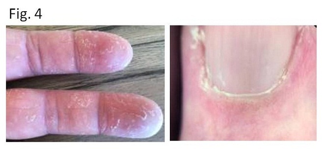
Anti-synthetase syndrome(ARS) is a chronic auto-immune condition or disease. The pathogenesis is not totally clarified. However, in anti-synthetase syndrome, autoimmune antibodies are producing against the synthetase enzyme called aminoacyl-tRNA synthetases that directed toward the attachment of particular amino acid to its transfer RNA (tRNA) (1, 2). The autoimmune antibodies arise after viral infections and patients might have a genetic proposition. They attack certain body cells, causing various symptoms; myositis; polyarthritis; interstitial lung disease; thickening and cracking of the hands (mechanics hands)(Fig. 4) ; Raynaud phenomenon (1, 2). Interstitial lung disease is known to precede to other symptoms (3, 4). In our case, interstitial lung disease was found alone and other symptoms were not, they might be appeared afterwards.
Pathological findings of interstitial pneumonia in anti-synthetase syndrome include organized alveolar spaces, alveolar collapse, fibrotic alveolar septum thickening and alveolar septum lymphocytes infiltration (5). These are histologic findings of non-specific interstitial pneumonia (NSIP) and organization pneumonia (OP). In short, NSIP is divided histologically into cellular NSIP and fibrotic NSIP. Cellular NSIP: lymphocytes infiltration to alveolar septum, fibrotic NSIP: thickened fibrosis of alveolar septum. Cryptogenic organizing pneumonia (COP) occurs approximately a half of OP (5). The histologic findings of COP are; alveolar space is occupied with organized tissue: occlusion of bronchiole by fibrosis. COP fibrosis can be absorbed and migration of COP lesions is often found on follow up chest CT, indicating COP fibrosis is absorbable. Interstitial pneumonia in anti-synthesis syndrome indicates a mixed pattern of COP and NSIP, implying interstitial pneumonia in anti-synthetase syndrome is not permanent but reversible, that is known to disappear after treatment (1-4).
The specific radiologic appearance of interstitial pneumonia of anti-synthetase syndrome is found to be ground glass opacification and/or consolidation in dorsal marginal area of both lower lobes rather than other lobes (1-5). As radiological findings, usual interstitial pneumonia (UIP) is found to be honey comb pattern in both lower lobes, especially susceptible to costophrenic angle and sub-plural area. NSIP is used as a term of pulmonary fibrosis except UIP. Namely, honey comb pattern is less at costophrenic angle or dorsal marginal area of both lower lobes but reticular pattern in NSIP. Approximately a half of NSIP is collagen vascular disease (4, 5). Meanwhile, COP is found to be patchy ground glass opacification in marginal area of a whole lung that can be migratory as time progress. Experimentally, COP is formed by T cell infiltration after disorder of pulmonary cell I. Radiologic findings of chronic eosinophilic pneumonia and anti-neutrophil cytoplasmic antibody (ANCA) pneumonia mimic COP that is formed by basement membrane disruption by eosinophilia infiltration.
Although honey comb pattern and reticular pattern at dorsal marginal area including costophrenic angle is found to be in UIP and NSIP, respectively, ground glass and/or consolidation at the same area might be specific in interstitial pneumonia of anti-synthetase syndrome. However, as time progress, chronic interstitial pneumonia of anti-synthesis syndrome turns to be reticular and honey comb pattern (8). Oral corticosteroids (prednisone) and immunosuppressive agents are the mainstay of treatment and very effective to improve symptoms and disappear chest shadow. Prompt diagnosis and appropriate treatment are favorable.
In our case, ground-glass and consolidation with neither honey comb nor reticular patterns were found at dorsal marginal area in both lower lobes, indicating a relatively acute and active phase of anti-synthetase syndrome.
【Summary】
A seventy fours-year-old male came to our hospital for persistent cough for two months and mild fever for five days despite antibiotics infusion. Although myositis and polyarthritis were not found, chest CT showed ground glass opacity and consolidation with neither honey comb pattern nor reticular pattern at the dorsal marginal area of both lower lobes which was considered specific CT findings of anti-synthetase (anti-aminoacyl-tRNA synthetases ARS) syndrome. We should keep in mind that histologic examination of interstitial pneumonia (ARS syndrome) reveals mixed pattern of COP and NSIP, but, different from typical radiological findings of COP and NSIP, CT shows ground glass opacity and consolidation at the dorsal marginal area of both lower lobes which are specific for acute type of this disease.
【References】
1.Antisynthetase syndrome. DermNet NZ. December 2014; http://dermnetnz.org/immune/antisynthetase.html.
2.Antisynthetase syndrome. Orphanet. May 2014; http://www.orpha.net/consor/cgi-bin/Disease_Search.php?lng=EN&data_id=8611.
3.Chatterjee S, Prayson R, Farver C.. Antisynthetase syndrome: not just an inflammatory myopathy. Cleve Clin J Med. October 2013; 80(10):655-666. https://www.ncbi.nlm.nih.gov/pubmed/24085811.
4.Koreeda Y, et al. Clinical and pathological findings of interstitial lung disease patients with anti-aminoacyl-tRNA synthetase autoantibodies. Intern Med. 2010;49(5):361-9. Epub 2010 Mar 1.
5.Friedman AW, et al. Interstitial lung disease with autoantibodies against aminoacyltRNAsynthetases in the absence of clinically apparent myositis. Semin Arthritis Rheum 1996;26: 459―467.
6.Naydour Q, et al. Anti-synthetase syndrome presenting as cryptogenic organizing pneumonia. Respir Med Case Rep. 2012; 6: 13–15. Published online 2012 Oct 6. doi: 10.1016/j.rmcr.2012.08.003 PMCID: PMC3920447
7.Thanuja SM, et al. Organizing pneumonia as the first manifestation of anti-synthetase syndrome. BMC Res Notes. 2016; 9: 290. Published online 2016 Jun 2. doi: 10.1186/s13104-016-2094-3 PMCID: PMC4890335
8.Debray MP, et al. Interstitial lung disease in anti-synthetase syndrome: initial and follow-up CT findings. Eur J Radiol. 2015 Mar;84(3):516-23. doi: 10.1016/j.ejrad.2014.11.026. Epub 2014 Dec Debray MP
2017.10.18
COPYRIGHT © SEICHOKAI YUJINKAI. ALL RIGHTS RESERVED.