

Clinical diagnosis
Case 9
【Clinical progress】
Day 2, she received surgical laparotomy which revealed the strangulation ileus, being given release of the incarcerated hernia, incision of the adhesion band and resection of a small part of the small bowel. Several days later, she discharged without any complication.
【Discussion】
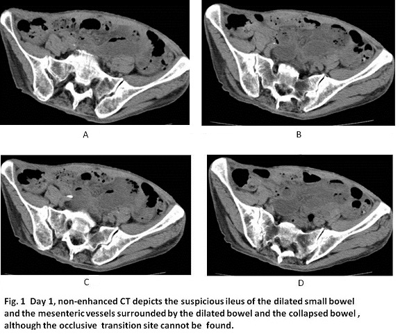
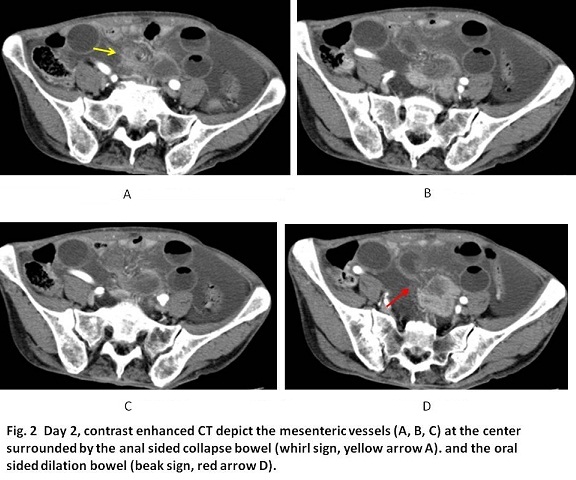
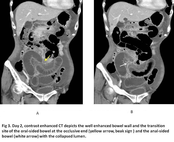
When ileus is encountered, it is one of the requirements of CT to differentiate between strangulation ileus and adhesive one or between laparotomy-applicable ileus and ileus tube-applicable one (1-5). The degree of strangulation ileus varies from torsion alone to torsion with ischemic necrosis. Pneumatosis of the bowel wall, thrombosed mesentery vessels (2, 4) and reduced enhanced bowel wall indicate high possibility of the the necrosis of the bowel (1, 5). Beak sign which implies the tapering occlusion of the bowel, and then, it can occur in both of anal sided and oral sided bowels. Beak sign is often associated with whirl sign which indicates the bowel encircles centering the mesenteric vessels. Whirl sign is the reliable CT finding of torsion (2, 4). The clue to find whirl sign is to check the mesenteric vessels and the encircling collapsed and dilated bowels around the mesenteric vessels (2, 3, 4). In our case, non-enhanced CT on Day 1 showed the difficulty to find the whirl sign and contrast-enhanced CT on Day 2 showed the well enhanced bowel wall and possible whirl sign at the only one level from a retrospective view point. Actually, the whirl sign was overlooked in the rough interpretation on Day 2. The detailed interpretation of CT findings should be required.
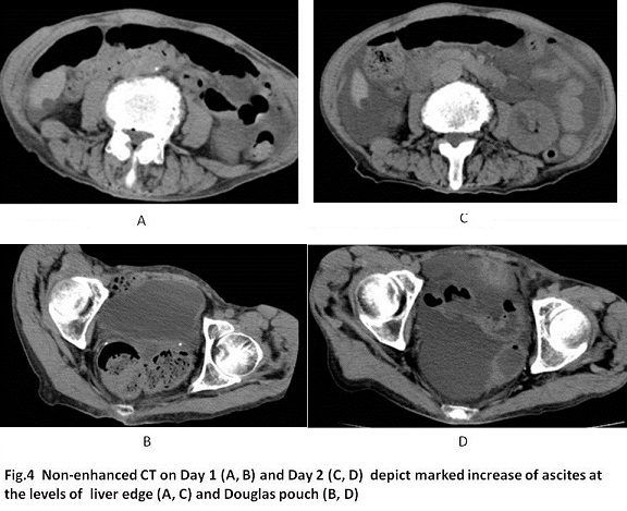
Meanwhile, the increasing ascites was found. When the mesentery torsion occurs, the portal vein tends to strangulate easily rather than the artery due to the thin vessel wall of the portal vein. The portal vein flow does not drain sufficiently, possibly leading to the mesentery edema, mesenteric fluid and ascites. Although the presence of ascites does not usually indicate the bowel necrosis, the increasing ascites implies at least the unchanged torsion, falling in the possible congestive necrosis sooner or later, which indicates the laparotomy-applicable ileus. Although the whirl sign was overlooked in our case, the increasing ascites was found on Day 2 and small intestinal resection was given. Then, it is considered that increasing ascites on CT was one of the determinant factors for laparotomy in our case. However, in general, increasing ascitis alone is not usually determinant because intraluminal pressure of the bowel without necrosis might happen to induce ascites.
【Summary】
A seventy five-year-old female suffering from the repeated vomits received CT on day 1 and day 2 which showed the increasing ascites, despite the difficult finding of whirl sign and reduced enhanced bowel wall, leading to laparotomy which revealed the strangulation ileus, and the patient was given a small part of intestinal resection . Several days later, the patient discharged without complication.
【References】
1.Ohira G, et al. Utility of arterial phase of dynamic CT for detection of intestinal ischemia associated with strangulation ileus. World J Radiol. 2012 28;4:450-454. doi: 10.4329/wjr.v4.i11.450.
2.Hayakawa K, et al. CT findings of small bowel strangulation: the importance of contrast enhancement. Emerg Radiol. 2013;20:3-9. doi: 10.1007/s10140-012-1070-z. Epub 2012 Aug 22.
3.Millet I, et al. Value of CT findings to predict surgical ischemia in small bowel obstruction: A systematic review and meta-analysis. Eur Radiol. 2015 Jun;25:1823-35. doi: 10.1007/s00330-014-3440-2. Epub 2015 Apr 8.
4.Mallo RD, et al. Computed tomography diagnosis of ischemia and complete obstruction in small bowel obstruction: a systematic review. J Gastrointest Surg. 2005 ;9 :690-4.
5.Geffroy Y et al. Increased unenhanced bowel-wall attenuation at multidetector CT is highly specific of ischemia complicating small-bowel obstruction. Radiology. 2014 ;270:159-167. doi: 10.1148/radiol.13122654. Epub 2013 Oct 28.
2016.06.08
COPYRIGHT © SEICHOKAI YUJINKAI. ALL RIGHTS RESERVED.