

Clinical diagnosis
Case 101
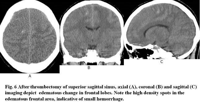
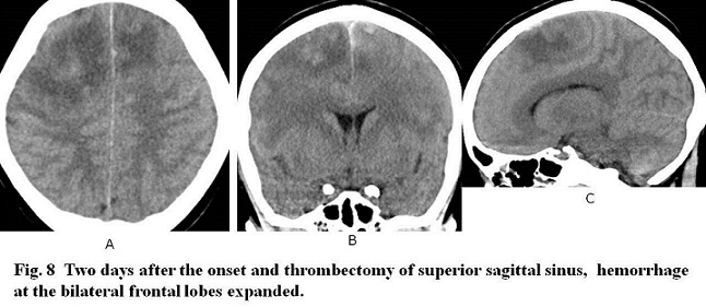
【Progress】Her symptoms of convulsion and headache worsened after MRI and CT. Angiography was conducted to confirm the occlusion of thrombosis at the superior sagittal sinus. Surgical thrombectomy was conducted soon after. CTs after the thrombectomy (Figs. 6, 7) and two days after (Fig. 8) were attempted to check the progress. They showed spotty hemorrhage and congestion at bilateral frontal lobes corresponded to areas draining into the anterior part of the sagittal sinus.

【Discussion】
Before investigating sinus thrombosis, we should refresh anatomy of the venous drainage of the brain. The main venous route is that superior sagittal sinus meets straight (rectal) sinus, forming confluent sinus, and then move to transverse sinus and sigmoid sinus, ending to internal jugular vein. Superior sagittal sinus communicates directly with inferior sagittal sinus via Trolard vein. Sagittal sinus get the drainage flow from cortical veins of frontal, parietal and occipital lobes. Then, thrombosis of superior sagittal sinus causes congestive hemorrhagic infarction in bilateral these lobes, and its affected areas depend on the site and the volume of sagittal sinus thrombus (1-6). In our case, anterior part of the sagittal sinus was thrombosed, leading to the congestive hemorrhagic infarction of bilateral frontal lobe and parietal lobe.
Straight sinus is formed by inferior sagittal sinus and Galen vein which is joined by bilateral internal cerebral veins. Internal cerebral vein gets the drainage flow from thalamo-striate veins. Then, it happens that thrombosis of internal cerebral vein, Galen vein or straight sinus causes congestive hemorrhage of bilateral basal ganglions or corpse callosum (1-6). Transverse (lateral) sinus get the drainage flow from Labbe vein. Then, it happens that thrombosis of Labbe vein or transverse sinus causes congestive hemorrhagic infarction of the temporal lobe (1-6).
Sigmoid sinus gets draining blood flow from superior and inferior petrosal veins originated from cavernous sinus. Cavernous sinus gets the draining blood flow from parietal lobe and temporal lobe. Thrombosis of cavernous sinus is yet to be reported.
Most systemic veins have venous valves, whereas cerebral venous sinuses do not have ones. Then, in coagulopathic conditions such as contraceptive drugs, puerperium and pregnancy, brain venous sinus is one of the susceptible veins for thrombosis. Adult patients with sinus thrombosis are women with the incidence of 75% (6). The sinus thrombosis occurs spontaneously with the incidence of 25% (1-3, 6). In our adult case, she was not pregnant and did not get hormonal drugs. The number of platelets is quite greater, 1500,000/mm3. Its cause was unfortunately unknown, probably spontaneous.
As radiologic findings, dense triangular sign of thrombosed superior sagittal sinus, cord sign of thrombosed transverse sinus or superior sagittal sinus,dense clot sign of thrombosed sinus on non-enhanced CT, empty delta sign of superior sagittal sinus on contrast-enhanced CT, non-flow void sign of sinus on MRI or high signal intensity of sinus on T1 weighted image are known. However, dense triangular sign and dense clot sign mimic normal variants (7-11). It is when to think of sinus thrombosis that hemorrhagic infarction occurs with cytogenic edema and vasogenic edema at bilateral parasagittal or bi-basal ganglion regions, temporal lobe, and peripheral cortex. In our case, dense clot sign (dense triangular sign), empty delta sign, cord sign of superior sagittal sinus, parasagittal edematous lesion including spotty hemorrhage were found on CT and MRI (Figs 1-5)(7-11).
As clinical manifestations, chronic headache (70%), motor or sensory deficit, seizures, drowsiness and mental change are listed (1-3). Our case experienced headache and motor deficit.
【Summary】
We present a twenty nine-year-old female suffering from vomiting, headache and left hemiparesis. Laboratory test revealed hemoglobin 3.6 g/dL and platelets 1500,000/mm3. Brain CT and MRI showed dense clot (triangular) sign & empty delta sign & cord sign and absent flow void sign of sagittal sinus & bilateral parasagittal edema with cytogenic edema, respectively. We should keep in mind that the image findings of possible thrombosis of sinus are bilateral hemorrhagic infarction with cytogenic edema and vasogenic edema at bilateral parasagittal or bi-basal ganglion regions, temporal lobe, and peripheral cortex. Further, CT and MRI show dense clot sign & empty delta sign and loss of flow void & high signal intensity on T1WI, respectively. Furthermore, thrombosis of sagittal sinus causes bilateral parasagittal hemorrhagic infarction, thrombosis of Labbe and transverse sinus causes hemorrhagic infarction of the temporal lobe, and thrombosis of internal cerebral vein, Galen vein and straight sinus cause hemorrhagic infarction of both basal ganglions.
【References】
1.Stam J . "Thrombosis of the cerebral veins and sinuses". N. Engl. J. Med. 2005; 352 (17): 1791–8. doi:10.1056/NEJMra042354. PMID 15858188
2.Cumurciuc R, et al. "Headache as the only neurological sign of cerebral venous thrombosis: a series of 17 cases". J. Neurol. Neurosurg. Psychiatry. 2005; 76 (8): 1084–7. doi:10.1136/jnnp.2004.056275. PMC 1739763 . PMID 16024884.
3.Einhäupl K, et al. "EFNS guideline on the treatment of cerebral venous and sinus thrombosis". Eur. J. Neurol. 2006; 13 (6): 553–9. doi:10.1111/j.1468-1331.2006.01398.x. PMID 16796579.
4.Ferro JM, et al. Prognosis of cerebral vein and dural sinus thrombosis: results of the International Study on Cerebral Vein and Dural Sinus Thrombosis (ISCVT). Stroke. 2004;35 (3): 664-70. doi:10.1161/01.STR.0000117571.76197.26 - Pubmed citation
5.Herrmann KA, et al. Thrombosis of the internal cerebral vein associated with transient unilateral thalamic edema: a case report and review of the literature. AJNR Am J
6.Ferro JM, et al. "Cerebral vein and dural sinus thrombosis in elderly patients". Stroke. 2005; 36 (9): 1927–32. doi:10.1161/01.STR.0000177894.05495.54. PMID 16100024.
7.Rodallec MH, Krainik A, Feydy A et-al. Cerebral venous thrombosis and multidetector CT angiography: tips and tricks. Radiographics. 2006;26 Suppl 1 : S5-18. doi:10.1148/rg.26si065505 - Pubmed citation
8.Poon CS, et al. Radiologic diagnosis of cerebral venous thrombosis: pictorial review. AJR Am J Roentgenol. 2007;189 (6_supplement): S64-75. doi:10.2214/AJR.07.7015 - Pubmed citation
9.Zeina AR, et al. Hyperdense cerebral sinus vein thrombosis on computed tomography. West J Emerg Med. 2011;11 (2): 217.
10.Lee Emil J. Y. “The Empty Delta Sign.” Radiology 2002; 224: 788-789. doi:10.1148/radiol.2243990978.
11.Virapongse C, et al.“The empty delta sign: frequency and significance in 76 cases of dural sinus thrombosis.”Radiology 162; 1987: 779-785. [Link]
2018.4.18