

Clinical diagnosis
Case 21
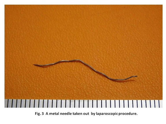
The laparoscopy revealed the presence of gastro-colic (great) omentum corresponded to the site of the skin marking. However, the foreign body could not be discovered at the skin marking site probably because of moving after air infusion into abdomen for laparoscopy. The foreign body was detectable under radio-fluoroscopy. When the great omentum was moved, radio-fluoroscopy revealed the foreign body simultaneously moved, indicating it existed in the greater omentum. The foreign body was eventually found out in the greater omentum and removed by laparoscopic procedure, which showed the metal needle(Fig. 3). It probably penetrated from the lumen of the digestive organ to the peritoneal lumen. There was no evidence of infectious contamination in the peritoneal space. Drainage and intra-peritoneal washing were not necessary. The patient was discharged eleven days later without any negative episodes
【Discussion】
The ingested foreign body mostly will pass through the entire organ uneventfully. As the perforated foreign body, sewing needles, dental plates, fish bones, toothpicks and metal needles were reported (1-6). The possible perforated site might be the less movable or angulated digestive segment such as the natural anatomical angularity of the lesser curvature in the stomach, C curve of the duodenum, the initial segment of jejunum beyond Treiz ligament, the ileum end and the recto-sigmoid junction (1, 2). In our case, a metal needle was found out in the great omentum. The great omentum is anatomically known to extend from the greater curvature of the stomach, passing in front of the small intestines and doubles back to ascend to the transverse colon before reaching to the posterior abdominal wall (2).Then, the metal needle of this case is supposed to originate from the stomach, the ligament of the Treiz, or the transverse colon.
When fish bones and toothpicks were perforated, peritonitis and/or abscess were documented to usually occur (5, 6). Meanwhile, in case of perforation with metal needle, inflammatory changes do not occur mostly (1-3). In our case, CT and laparoscopy revealed no evidence of inflammatory contamination. The inorganic substance of the metal needle is considered not to trigger bacterial infection.
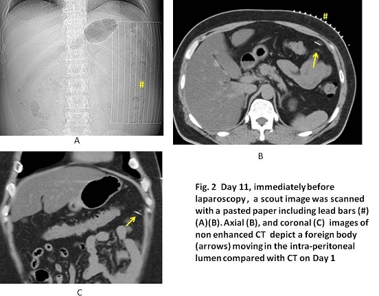
Before laparoscopy, CT using grid-like bar on the surface of the abdomen was conducted to identify the exact site of the foreign body and to make the skin mark. However, it was useless because of air infusion for laparoscopy inducing the movement of the great omentum. Meanwhile, radio-fluoroscopy was useful to detect the foreign body of metal component during laparoscopy (2). In case of something like fish bone with less metal component, laparotomy might be followed by laparoscopic trial because of the difficult identification by radio-fluoroscopy .
【Summary】
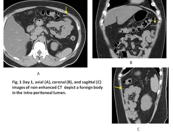
We present a case with a foreign body dected on CT. He did not remember to ingest something strange accidentally. As he was a chef and had many chances to eat fish bones, the foreign body was suspected at first to be something related with fish. Laparoscopy revealed that the foreign body was a metal needle which was found in the great omentum with no evidence of inflammatory contamination.
【References】
1.Aarabi S, et al. Noningested intraperitoneal foreign body causing chronic abdominal pain: a role for laparoscopy in the diagnosis. J Pediatr Surg. 2012;47:e15-7. doi: 10.1016/j.jpedsurg.2011.10.052.
2.Bulbuloglu E, et al. Laparoscopic removal of a swallowed sewing needle that migrated into the greater omentum without clinical evidence. J Laparoendosc Adv Surg Tech A. 2005;15:66-69.
3.Varetto L, et al. Intra-abdominal needle: medical malpractice? Forensic Sci Int. 2009 ;191:e11-3. doi: 10.1016/j.forsciint.2009.07.011. Epub 2009 Aug 13.
4.Kendra Klein, et al. Transluminal migration of ingested foreign body without peritonitis. Journal of Pediatric Surgery 2012; 47: 788-791
5.Venkatesh SH, et al. CT findings of accidental fish bone ingestion and its complications. Diagn Interv Radiol. 2016 Mar; 22(2): 156–160. Published online 2015 Dec 29. doi: 10.5152/dir.2015.15187
6.Reginelli A, et al. Computed Tomographic Detection of Toothpick Perforation of the Jejunum: Case Report and Review of the Literature. Radiology Case Reports.2007; 2: 17-21.
2016.8.31
COPYRIGHT © SEICHOKAI YUJINKAI. ALL RIGHTS RESERVED.