

Clinical diagnosis
Case 49
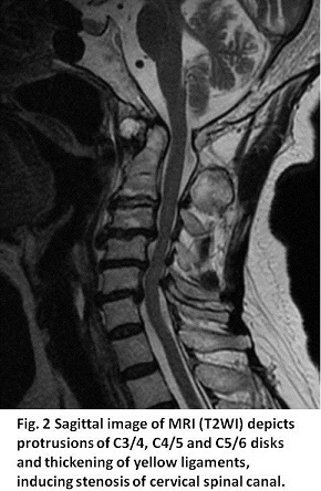
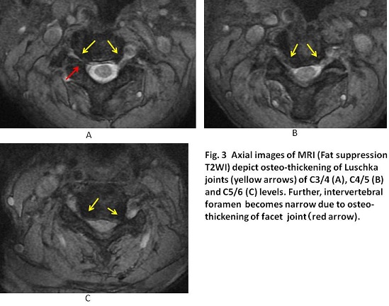
Based on MRI findings described in figure legends of Figs 2 and 3, imaging diagnosis was made; Luschka arthritis: spinal canal stenosis due to protrusion of C3/4, C4/5 and C5/6 disks and thickening of yellow ligament.
【Discussion】
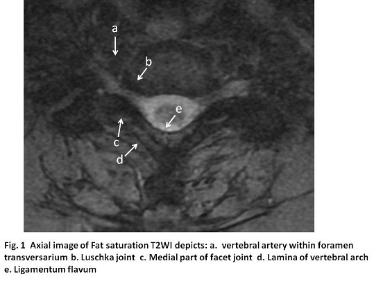
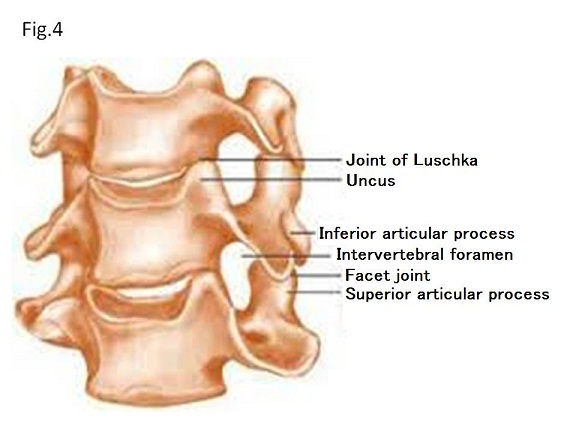
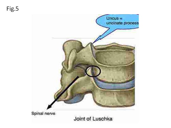
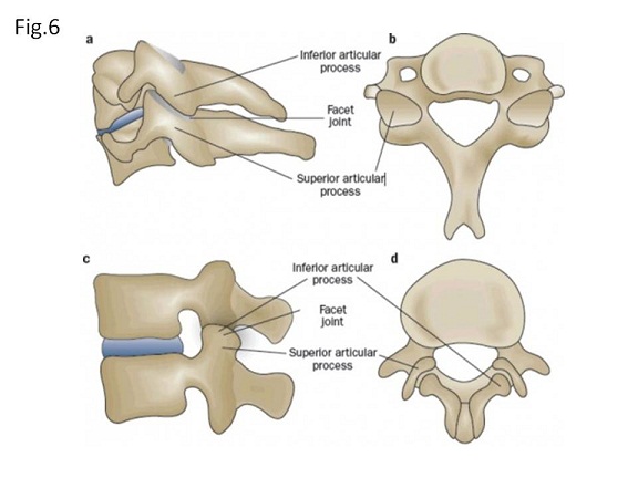
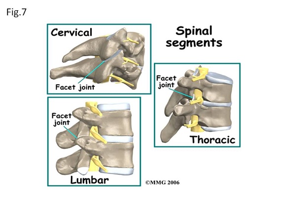
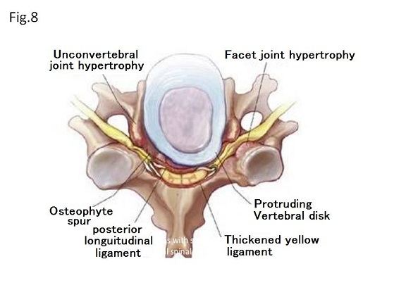
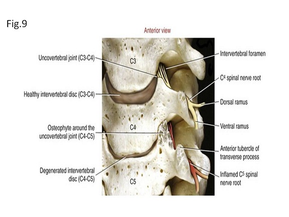
Luschka joint is formed between an uncus which projects from the lower vertebra and lateral aspect (shallow concave) of the upper vertebral body (1, 2, 3)(Fig. 4). Luschka joint forms bilaterally like a cat ear shape and only found in C2/3 to C6/7 levels (C1 and C2 own no uncus). Although these joints allow flexion and extension, they probably function to limit lateral movements of the cervical vertebrae and stable C3 to C7 together while C1 and C2 function to rotate head. In fact, Luschka joints do not exist in thoracic spine and lumbar spine (1, 2). Luschka joint is also called uncovertebral joint. The important factor is that this joint forms the anterior border of the intervertebral foramen for nerve roots. When cervical disks become drying and shrinking, uncus starts rubbing on the vertebral body and induces degenerative joint change: osteophytes (spurs) growing. Spurs start compress the nerve root, causing radicular pain. Our patient suffered from bilateral hand numbness, and MRI showed the bony thickening of anterior border of the intervertebral foramen (Fig. 3). Facet joint is termed intervertebral joint or zygapophyseal joint (3, 4). Two facet joints exist at every level of the vertebral column. Facet joint is formed between inferior articular process from the upper vertebra and superior articular process from the inferior vertebra. The paired facet joint and the intervertebral disk are called ‘three-joint complex’. They functions together to transfer loads, and to guide and constrain motions in the spine (3, 4). Interestingly, although the facet joints are aligned to allow flexion and extension, the facet joints runs anterior-superior to posterior-inferior in the cervical facet joint (Figs. 6, 7), frontal in the thoracic facet joint and sagittal in the lumbar spine (Fig. 7). When vertebral disk is getting dry-out and shrink, the burden of the paired facet is getting increased, inducing facet joint osteoarthritis (Figs. 8, 9); thickening and deformity of superior and/or inferior articular processes; narrowing of the facet joint space; subarticular bone erosion, subchondral cysts and osteophyte formation, and bone marrow erosion. The nerve root runs directly under the facet joint and as the facet joint becomes narrow due to bony thickening, it possibly pinches the nerve root. It is important to recognize that this joint forms the posterior border of the intervertebral foramen for nerve roots (Figs. 8, 9). In our case, the bony- thickening from the posterior border was found in Fig. 3A.
【Summary】
We present a seventy nine-year-old female with Luschka joint arthritis and facet joint arthritis. We should keep in mind that on axial images of cervical spine, the anterior border of the intervertebral foramen for nerve roots composes of Luschka joint, while the posterior border of the intervertebral foramen composes of medial part of facet joint. Cervical spine has the distinctive characters of having C1 and C2 for specific configuration, Luschka joints (C3 –C7) and foramen transversarium in transverse process (C1 – C7).
【References】
1.Bryan O'Young, et al. Physical medicine and rehabilitation secrets. Elsevier Health Sciences. pp. 46–. ISBN 978-1-56053-437-2. Retrieved 19 May 2011.
2.David J. Magee. Orthopedic physical assessment. Elsevier Health Sciences. pp. 134–. ISBN 978-0-7216-0571-5. Retrieved 19 May 2011.
3.Hubert Luschka . 1858 Die Halbgelenke des menschlichen Körpers: Mit 6 Kupfertafeln. Ge. Reimer. Retrieved 19 May 2011.
4.Morishita K, et al. Hypertrophic change of facet joint in the cervical spine. Med Sci Monit. 2008 Feb;14(2):CR62-64.
5.Alfred C. Gellhorn, et al. Osteoarthritis of the spine: the facet joints.Nat Rev Rheumatol. 2013 Apr; 9(4): 216–224. Published online 2012 Nov 13. doi: 10.1038/nrrheum.2012.199
2017.4.5
COPYRIGHT © SEICHOKAI YUJINKAI. ALL RIGHTS RESERVED.