

Clinical diagnosis
Case 5
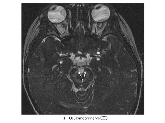
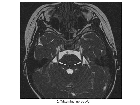
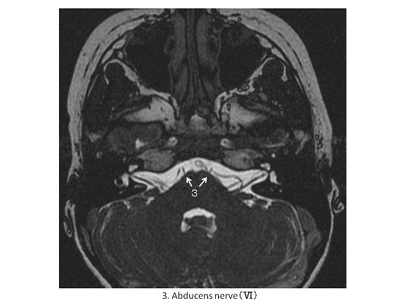
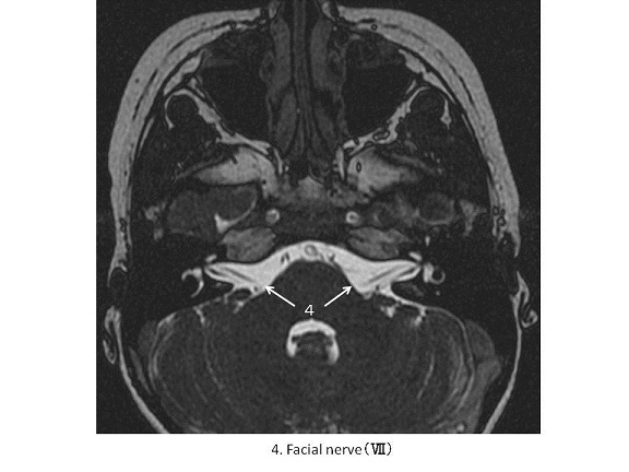
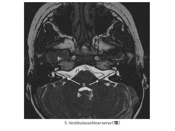
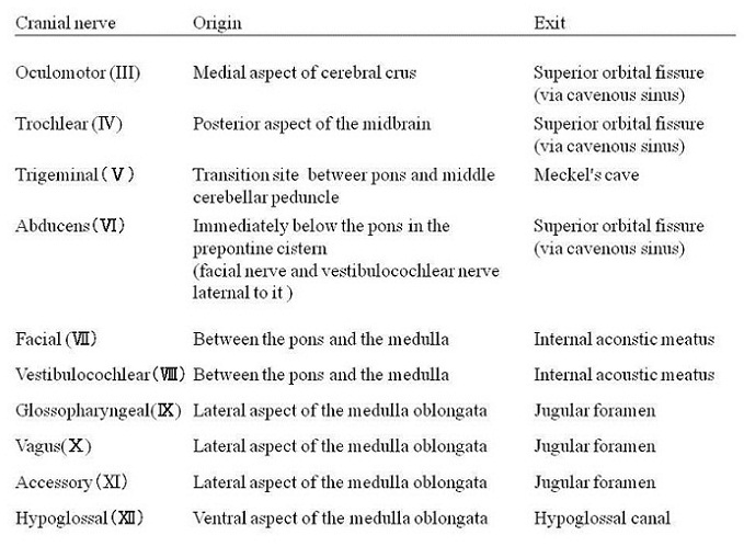
In order to visualize cranial nerves, balanced SSFP (steady-state free precession) is used as an ultra-high speed sequence of MRI. The signal intensity of balanced SSPF is corresponded to the ratio of T2/T1 (1). Because the ratio of T2/T1 is the greatest in free water and the secondary greatest in, two characteristics of the SSFP image are listed; free water is visualized with high signal intensity: vascular lumen is also visualized with high signal intensity (1). Then, cranial nerves resulted in low signal intensity differentiating from high signal intensity of cerebral-spinal fluid and vessels lumen. The facial nerve, the seventh paired cranial nerve (CN VII) arises in the pons, and it originally begins as two roots; a large motor root innervates the muscle of facial expression: a small sensory root provides the function of taste sensation to the anterior 2/3 of the tongue and parasympathetic excretion of the glands of the face and neck including the submandibular, sublingual, nasal, lacrimal and pharyngeal glands. The two roots fuse together and travels to the internal acoustic pore (meatus) situated in the petrous part of the temporal bone, together with the vetibulocochlear nerve (CNVIII).
【Summary】
Cranial nerves can be identified using a balanced SSFP sequence of MRI which depicts a facial nerve at the level of internal acoustic pore (meatus), travelling with the vetibulocochlear nerve (CNVIII).
【References】
1.Araki T. Keeteibann MRI kannzennkaisetu (Japanese) pp 250-251 Syujunsya 2008
2.Gupta, Sachin; Francine Mends; Mari Hagiwara; Girish Fatterpekar; Pamela C. Roehm (2013). "Imaging the Facial Nerve: A Contemporary Review". Radiology Research and Practice 2013: –248039. doi:10.1155/2013/248039. ISSN 2090-1941. Retrieved 2015-02-07.
3.Jump up^ PhD, Richard S. Snell MD (2011). Clinical Anatomy by Regions (Ninth ed.). Philadelphia, Pa.; London: LWW. ISBN 9781451110326.
4.Jump up^ Singh, Vishram. Textbook of Clinical Neuroanatomy (2nd ed.). p.104.
2016.05.11
COPYRIGHT © SEICHOKAI YUJINKAI. ALL RIGHTS RESERVED.