

Clinical diagnosis
Case 61
Our orthopedist advised him not to play handball but keep rest. He was treated by brace covering his right forefoot. He did not want non-steroidal anti-inflammatory drugs (NSAIDs).
【Progress】
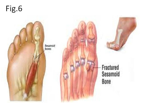
Sesamoids (or sesamoid bones) are focal ossification areas within tendons as they pass over joints (1-3). The term of “sesamoid bone” seemed to be named as an alternative of a small bone or a round-shaped bone probably looking like a sesame seed. A sesamoid bone usually exists at the site of the joint and embedded in the tendon where it crosses over a joint (1-3). The largest sesamoid bone is a knee cap or patella. A hyoid bone does not belong to a sesamoid bone because it forms from cartilage and exists not relevant with a joint. The sesamoid bone is not present at birth. Ossification of the sesamoid bone of the thumb presents at the age of 10 to 12 (4).
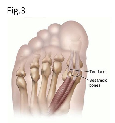
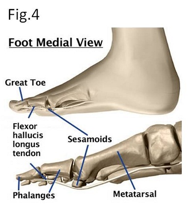
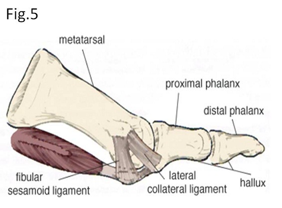
Sesamoids bones act like fulcrum points of tendon and function such as pulleys. In short, a sesamoid bone makes the tendon or the ligament goes slightly further away from the joint, inducing to increase the smooth movement of the tendon and to inhibit the tendon from flattering into the joint as tension increases (Figs 3, 4). In case of the forefoot, sesamoid bones form the bump at the underside of the big toe (Figs 3, 4). The toe flexor muscle (flexor hacillus longus tendon) contacts sesamoid bone and pass underneath the first metacarpo-pharyngeal (MP) joint (Fig. 5). When this muscle bends the big toe, the sesamoid bone functions as a fulcrum point for the flexor muscle with gliding over MP joint, helps to absorb pressure from under the foot during standing and walking, and eases friction between MP joint and toe flexor muscle when the big toe moves (Figs 3 – 5).
【Summary】
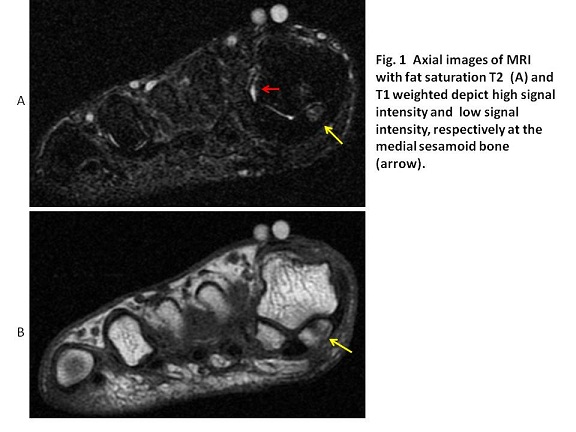
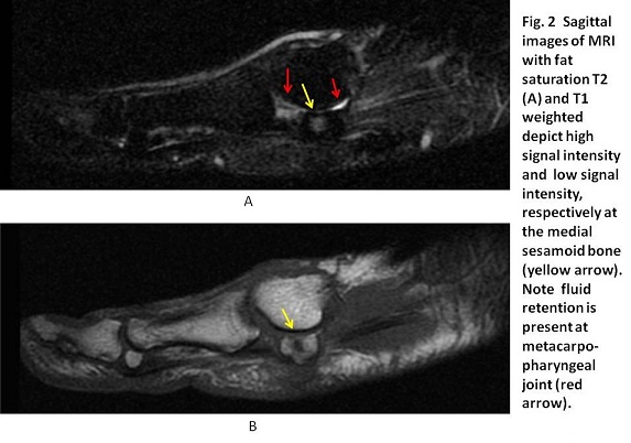
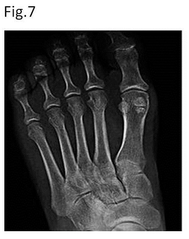
We present a sixty nine-year-old male with stress fracture of right medial sesamoid bone which was visualized on fat-suppression T2WI and T1WI. He was an athlete of handball that long-standing and repeated weight loading to the big toe was considered to cause the sesamoid bone injury. We should keep in mind that sesamoid bones is present at the joint and is embedded in the tendon and act such as fulcrum points of tendon and pulleys for the purpose of the tendon going slightly further away from the joint, inducing to increase the movement of the tendon and to inhibit the tendon from flattering into the joint as tension increases, inducing the flexor muscle glides over MP joint smoothly and eases friction between MP joint and toe flexor muscle when the big toe moves.
【References】
1.Boike A, Schnirring Judge M, McMillin S. Sesamoid disorders of the first metatarsophalangeal joint. Clin Podiatry Med Surg. 2011;28(2):269–85. [PubMed]
2.Lee DK, Mulder GD, Schwartz AK. Hallux, sesamoid, and first metatarsal injuries. Clin Podiatry Med Surg. 2011;28(1):43–56. [PubMed]
3.Cohen BE. Hallux sesamoid disorders. Foot Ankle Clin. 2009;14(1):91–104. [PubMed]
4.Chaumoître K, et al. Value of the sesamoid bone of the thumb in the determination of bone age J Radiol. 2008 Dec;89(12):1921-4.
5.Biedert R, Hintermann B. Stress fractures of the medial great toe sesamoids in athletes. Foot Ankle Int. 2003;24(2):137–41. [PubMed]
6.Parra E. Stress fractures of the sesamoid. Clin Orthop. 1960;18:281–85.
7.Kalantari BN, Seeger LL, Motamedi K, Chow K. Accessory ossicles and sesamoid bones: Spectrum of pathology and imaging evaluation. Appl Radiol. 2007;36(10):28-37. Appl Radiol (full text)
8.Dobas DC, Silver MD. The frequency of the bipartite sesamoids of the first metatarsophalangeal joint. J Am Podiatry Assoc. 1977;67(12):880–82. [PubMed]
9.Frankel J, Harrington J. Symptomatic bipartite sesamoids. J Foot Surg. 1990;29:318–23. [PubMed]
10.Blundell CM, Nicholson P, Blackney MW. Percutaneous screw fixation for fractures of the sesamoid bones of the hallux. J Bone Joint Surg Br. 2002;84(8):1138–41
2017.7.5
COPYRIGHT © SEICHOKAI YUJINKAI. ALL RIGHTS RESERVED.