

Clinical diagnosis
Case 63
【Progress】
After imaging diagnosis of lateral discoid meniscus, she came back to the local orthopedist. Thereafter, we have no information about treatment of her right knee.
【Discussion】
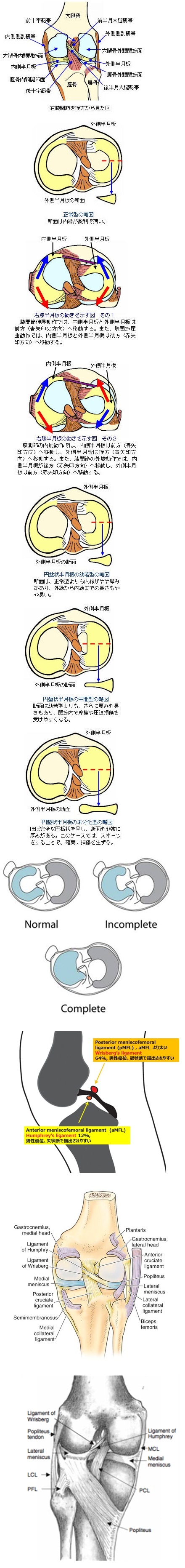
A normal knee meniscus composes of fibrocartilage and forms like a crescent configuration with much opening inside space. It basically functions to act as a stabilizer with ligaments, spacer to avoid friction between the femur and tibia and shock absorber that lower stress to the articular cartilage (1, 2).
A discoid meniscus forms like a half moon to full moon configuration with closing inside pace. A discoid meniscus is a normal variant and occurs in the lateral meniscus with the incidence of 0.8 – 3 % of the population (2 - 6). A lateral discoid meniscus is categorized into three types (1, 7); Type I, a complete disk of meniscus tissue completely covering the tibial plateau; Type II, that incompletely covering the tibial plateau; Type III, Wrisberg variant even if the meniscus have a normal configuration, lateral meniscus lacks coronary ligament which attaches horn to lateral joint capsule and/or its posterior horn attachment such as posterior superior and/or posterior inferior popliteomeniscal fascicle with popliteous tendon, symptomatic of locking, posterior horn snapping and mobile occasionally subluxing into the joint (2, 8). Type I is the most common. Lateral discoid meniscus with Types I and II are more susceptive of meniscus shearing than normal meniscus because of the greater coverage between tibia and femur joint (9 – 12).
In our case, her symptoms are knee pain and snapping (clicking). MRI showed a lateral discoid meniscus covering the total tibial plateau space with high signal intensity in its inside, implying Type I discoid meniscus and suspecting minor injury of discoid meniscus (Figs 1, 2, 4). Further, MRI showed high signal intensity corresponded to lateral and posterior interpose space surrounding discoid meniscus in T2WI, indicating the weak attachment to the joint capsule (Figs 2, 3). Both attachment of posterior horn of lateral discoid meniscus to popliteal tendon via popliteomeniscal fascicle and attachment of lateral discoid meniscus and coronary ligament seemed to be weaker rather than the opposite normal medial meniscus (Figs 4, 7). It implied the reason of occurrence of snapping during flexion or extension because snapping is considered to occur in friction of ligament to bone or friction of meniscus to tendon. Namely, she had Type I discoid meniscus and a factor of Wrisberg variant (Type III).
Furthermore, it is known that meniscofemoral ligament has two parts of anterior and posterior meniscofemoral ligament called Hamphry ligament and Wrisberg ligament, respectively. Wrisberg ligament connects between medial condyle of femur and posterior horn (2, 8). Discoid meniscus has only one Wrisberg ligament, indicating unstable posterior horn. Wrisberg ligament is thick and high in patients with a complete discoid lateral meniscus (2, 8) . In our case, thick posterior meniscofemoral (Wrisberg) ligament was present (Fig. 3A) and anterior meniscofemoral (Hamphry) ligament was not identified, probably causing unstable knee joint and making a sound of snapping or clicking of posterior horn during extension and flexion.
【Summary】
We present a thirteen-year-old girl suffering from right knee pain and snapping (clicking) for several months. Knee MRI showed lateral discoid meniscus with minor injury probably causing knee pain and thick posterior meniscofemoral (Wrisberg) ligament characteristic of Type I. Further, it showed weak attachments between posterior horn and popliteal tendon via popliteomeniscal fascicle, and between lateral discoid meniscus and coronary ligament probably causing snapping. These findings and illness history suggests her lateral discoid meniscus include Type I plus a factor of Type III.
【References】
1.Clark CR, Ogden JA. Development of the menisci of the human knee joint: Morphologic changes and their potential role in childhood meniscal injury. J Bone Joint Surg Am. 1983;65:538-547.
2.Singh, K et al. MRI Appearance of Wrisberg Variant of Discoid Lateral Meniscus American Journal of Roentgenology. 2006;187: 384-387. 10.2214/AJR.04.1785
3.Neuschwander DC, et al. Lateral meniscal variant with absence of the posterior coronary ligament. J Bone Joint Surg Am 1992; 74:1186-1190 [Medline]
4.Helms CA. The meniscus: recent advances in MR imaging of the knee. AJR 2002; 179:1115-1122 [Abstract]
5.Silverman J, et al. Discoid menisci of the knee: MR imaging appearance. Radiology 1989; 173:351-354 [Medline]
6.Rohren EM, et al. Discoid lateral meniscus and the frequency of meniscal tears. Skeletal Radiol 2001; 30:316-320 [CrossRef] [Medline]
7.Kelly BT, et al. Discoid lateral meniscus in children. Curr Opin Pediatr 2002; 14:54-61 [CrossRef] [Medline]
8.Watanabe M, Takada S, Ikeuchi H. Atlas of Arthroscopy. Tokyo, Japan:Igaku-Shoin;1969.
9.Kim EY, et al. Atypically thick and high location of the Wrisberg ligament in patients with a complete lateral discoid meniscus. Skeletal Radiol. 2008;37:827-833.
10.Kaplan EB. Discoid lateral meniscus of the knee joint: Nature, mechanism, and operative treatment. J Bone Joint Surg Am. 1957;39:77-87.
11.Connolly B, Babyn PS, Wright JG, Thorner PS. Discoid meniscus in children: Magnetic resonance imaging characteristics. Can Assoc Radiol J. 1996;47:347-354.
12.Youderian A, Chmell S, Stull MA. Discoid lateral meniscus. Applied Radiology. 2008;37:30-32.
13.Samoto N, Kozuma M, Tokuhisa T, Kobayashi K. Diagnosis of discoid lateral meniscus of the knee on MR imaging. Magn Reson Imaging. 2002;20:59-64.
2017.7.19