

Diagnosis
Case 68
【Progress】Two days later, bronchoscope and bronchoalveolar lavage (BAL) revealed the rate of eosinophilic cells of the total white blood cells was 31.5% Laboratory tests of the whole blood at that time revealed PO2 40.1%(83 -106), CRP 5.02 mg/dL, white blood cells 8100 / mm3 , eosinophils 30. 4 %, non-specific IgE 4514 IU/mL (14 - 400). Steroid administration was administered, inducing the improvement of her respiratory symptoms. One month later, she was discharged without complications.
【Discussion】
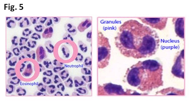
Of major three cells of mast cells, basophils and eosinophils in charge of allergic reaction, mast cells living in the tissue are related to immediate allergic response, basophils living in whole blood are related to in late allergic response and eosinophils living mainly in gastrointestinal and respiratory tracts are related to excrete their granules for cyto-toxicity attacking organisms such as bacillus and parasites (1). Eosinophils contain approximately 200 granules in the cytoplasm (Fig. 5)(1). They are formed in the bone marrow and travel in the vessels for around 10 hours before they arrive at the destination (1-5). The life span of eosinophils ranges from 2 to 5 days (1-5). Peripheral eosinophils counts less than 500/mm3 are normal. Eosinophils own various functions, moving inflamed area, trapping substance, killing cells, anti-parasitic and bactericidal activity, participating immediate allergic reaction and modulating inflammatory process (1-7).
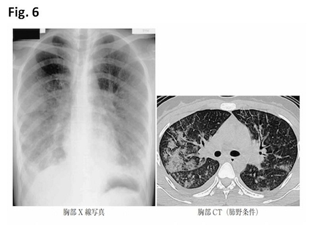
Eosinophils function mainly to be helpful but can occasionally be harmful. When eosinophils respond and infiltrations to the target, interstitial edema always occurs via excretion of leukotrien C4 and platelet activating factor from eosinophils which accelerate vessel permeability. In short, interstitial edema is critically found in pathological specimen (8). Chest CT findings of acute eosinophilic pneumonia (AEP) reflect interstitial edema; pulmonary edema; proximal bronchovascular bundle thickening; consolidation surrounding the primary bronchus: pleural effusion (Fig. 6). In our case, chest radiograph and chest CT showed the mimic images such as cardiac failure of butterfly pattern consolidation and pleural effusion (Figs. 1 – 4)(8).
Etiology causing AEP is fully unknown. However, it is well known that AEP occurs after cigarette smoking for the first time or following a period of smoking cessation (9-11). AEP can be caused by passive smoking and the inhalation of smoke from fireworks or bonfire (10-12). Drugs and vaccination also have been reported to be causative of AEP. Eosinophils are under control of T-lymphocytes. The activation of T-lymphocytes through cytokines, especially interleukin 5 is the main growth factor for maturation of eosinophils in bone marrow and migrate the habitant area via blood flow (3). In our case, it is considered that allergic reaction to drugs for Helicobacter Pylori caused edema of eyelids and foot, and AEP.
As diseases termed ‘eosinophilic’ except AEP, chronic eosinophilic pneumonia (similar CT image as cryptogenic organizing pneumonia), eosinopjilic sinusitis (mainly seen in opacification of ethmoid sinus), eosinophilic granuloma with polyangitis (similar CT image as Aspergillosis) (13) and eosinophilic gastroenteritis are listed. The term of “eosinophilic” is named based on microscopic eosinophilic infiltration, indicative of the immune response or disorder. Although it is unknown whether eosinophils are playing a main role or an assistant role in these diseases, eosinophils seem to work the leading part in causing AEP.
A diagnosis of AEP is made of elevation of eosinophils (greater than 500/mm3) in laboratory test and bronchoalveolar lavage (BAL)(greater than 25 %)(14). In our case, laboratory test of whole blood revealed the number of eosinophils on Day 1 and Day 3 was 977 and 2430/mm3 and BAL revealed rate of eosinophils was 30.4%.
【Summary】
We present an eighty nine-year-old female with AEP. After cessation of drugs for Helicobacter Pylori, she found edema of eyelids, swollen foot, fever and dyspnea. Chest radiograph and chest CT showed pleural effusion and consolidation and butter fly pattern usually found in cardiac failure: a bilateral consolidation pattern was found along with proximal bronchial trees. Laboratory test and BAL revealed greater number of eosinophils, indicative to fulfill the criteria of AFP. We should keep in mind that eosinophils cause the permeability of vessels, inducing pleural effusion and pulmonary edema such found in cardiac heart failure. Although it is known that AEP occurs after cigarette smoking for the first time or following a period of smoking cessation, drugs can also be causative of AEP. Peripheral eosinophils counts less than 500/mm3 are normal.
【References】
1.Kelly D. Stone, et, al. IgE, Mast Cells, Basophils, and EosinophilsJ Allergy Clin Immunol. 2010 Feb; 125(2 Suppl 2): S73–S80. doi: 10.1016/j.jaci.2009.11.017
2.Badesch DB, et al. Acute eosinophilic pneumonia: a hypersensitivity phenomenon? Am Rev Respir Dis 1989; 139:249.
3.Allen JN, et al. Acute eosinophilic pneumonia as a reversible cause of noninfectious respiratory failure. N Engl J Med 1989; 321:569.
4.Arrese M, et al. Eosinophilic pneumonia causing severe respiratory insufficiency. Clinical case. Rev Med Chil 1991; 119:299.
5.Nishio M, et al. Idiopathic acute eosinophilic pneumonia. Intern Med 1992; 31:1139.
6.Slabbynck H, et al. Dyspnea, fever, and eosinophilia. Chest 1993; 104:585.
7.Philit F, et al. Idiopathic acute eosinophilic pneumonia: a study of 22 patients. Am J Respir Crit Care Med 2002; 166:1235.
8.Mochimaru H, et al. Clinicopathological differences between acute and chronic eosinophilic pneumonia. Respirology. 2005 Jan;10(1):76-85.
9.Pope-Harman AL, et al. Acute eosinophilic pneumonia. A summary of 15 cases and review of the literature. Medicine (Baltimore) 1996; 75:334.
10.Miki K, et al. Early-phase neutrophilia in cigarette smoke-induced acute eosinophilic pneumonia. Intern Med 2003; 42:839.
11.Chung MK, et al. Acute eosinophilic pneumonia following secondhand cigarette smoke exposure. Tuberc Respir Dis (Seoul) 2014; 76:188.
12.Hirai K, et al. Acute eosinophilic pneumonia associated with smoke from fireworks. Intern Med 2000; 39:401.
13.Johkoh T, et al. Eosinophilic lung diseases: diagnostic accuracy of thin-section CT in 111 patients. Radiology. 2000;216:773–780.[PubMed]
14.Katoh S, et al. Elevated interleukin-18 levels in bronchoalveolar lavage fluid of patients with eosinophilic pneumonia. Allergy. 2004;59:850–856. [PubMed]
2017.8.23
COPYRIGHT © SEICHOKAI YUJINKAI. ALL RIGHTS RESERVED.