

Our treatment
Case 99
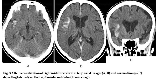
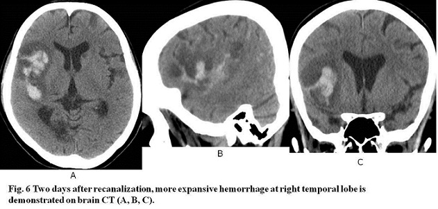
【Progress】Immediately after the successful retrieval of embolus, his Glasgow coma scale elevated from E4V4M6 to E4V5M6. However, brain CT after the procedure showed hemorrhage at the left insula, and two days later, brain CT showed the extension of the hemorrhage to the left whole temporal lobe.
【Discussion】
Of the four lobes of brain, a pair of temporal lobe are largest. The temporal lobes are involved in eight functions according to Kolb, B and Wishaw(1), IQ: 1) auditory sensation and perception 2) visual perception 3) selective attention of auditory and visual input 4) organization and categorization of verbal material 5) language comprehension 6) long-term memory 7) preserve of personality and behavior 8) preserve of sexual behavior. Wernicke’s area in the temporal lobe is vital to language comprehension and speech production.
Insula is situated inside of the Sylvian (lateral) fissure and lies under operculum (like a lid) which is formed by the portions of frontal lobe, temporal lobe and parietal lobe. Insula itself is called the fifth lobe or a part of the temporal lobe. Insula gets sensory signals from thalamus and connects to amygdala and temporal lobe, frontal lobe and parietal lobe (2). The function of insula was least known until 21st century. Through functional MRI imaging, function of insula is getting clarified. Insula is involved in remarkable functions: perception of pain, basic emotions (joy, happiness, anger, disgust), addiction and desire, reading emotions and social cues, motor control and above all, the sense (our perception) of ourselves. In short, of the functions of the temporal lobe described above, insula is involved in functions of preserve of personality and behavior, and preserve to sexual behavior (3, 4).
The middle cerebral artery arises from internal carotid artery and is one of the two main terminal branches (the other one: anterior cerebral artery).The middle cerebral artery is categorized into 4 parts; M1 the sphenoid (horizontal) segment branches a number of perforating arteries including lateral lenticulostriate artery which is called hemorrhagic artery, and internal lenticulostriate artery; M2 the insular segment; M3 the opercular segment exists in the Sylvian fissure: M4 cortical segment begins from Sylvian fissure and extends to the cortex.
The insula is irrigated by perforating branch of M2 and susceptible to ischemia because of the least development of collaterals from the other cerebral artery (5). When middle cerebral artery is occluded, insular ribbon sign is indicative of subtle early CT sign of infarction in the territory of the middle artery. Insular ribbon sign refers to hypodensity and swelling of the insular cortex (5, 6).
In our case, brain CT approximately eight hours after the onset of brain embolism showed insular ribbon sign. After catheter retrieval of embolism, hemorrhagic infarction occurred first at the right insula followed by the expansion to the territory of M2 or more peripheral (6).
The damage of insula is known to cause progressive expressive aphasia, which is the deterioration of verbal function to lose the ability to communicate with others fluently. The symptoms of language problem worsened as time progress in our patient.
【Summary】
We present a seventy three-year-old male suffering from sudden disorders of speech articulation and motility of left upper and lower extremities. Electrocardiogram revealed cardiac fibrillation. Brain CT showed positive insular ribbon sign and high density of the branches (M2 & M3) of the right middle cerebral artery, indicative of acute infarction. MRI & MRA showed occlusion of the right middle cerebral artery. After catheter retrieval of the emboli, hemorrhagic infarction was found at the insula and the temporal cortex. We should keep in mind that CT findings for early brain infarction in the region of the middle cerebral artery: a hyperdense segment of the middle cerebral artery, insula ribbon sign, and hypoattenuation of the ischemic region such as basal ganglion that is not found in our case. Further, the middle cerebral artery is categorized into M1 the sphenoid (horizontal) segment, M2 the insular segment, M3 the opercular segment and M4 cortical segment. Insula functions perception of pain, basic emotions (joy, happiness, anger, disgust), addiction and desire, reading emotions and social cues, motor control and above all, the sense (our perception) of ourselves, indicative of the characteristic of ourselves.
【References】
1.Kolb, B. and Wishaw, I.Q. (1990) Fundamentals of Human Neuropsychology. W. H. Freeman and Company, New York.
2.Nieuwenhuys R. The insular cortex: a review. Prog. Brain Res. 2012;195: 123-63. doi:10.1016/B978-0-444-53860-4.00007-6 - Pubmed citation
3.Sara W. et al. (November 2005). Meditation experience is associated with increased cortical thickness. NeuroReport 2005;16: 1893-1897.
4.Ogino Y, et al. "Inner experience of pain: imagination of pain while viewing images showing painful events forms subjective pain representation in human brain". Cereb. Cortex. 2007 17 (5): 1139–46. doi:10.1093/cercor/bhl023. PMID 16855007.
5.Delion M, Mercier P. Microanatomical study of the insular perforating arteries. Acta neurochirurgica. 156 (10): 1991-7; discussion 1997-8. doi:10.1007/s00701-014-2167-9 - Pubmed
6.Srinivasan A, Goyal M, Al Azri F, Lum C. State-of-the-art imaging of acute stroke. Radiographics : a review publication of the Radiological Society of North America, Inc. 26 Suppl 1: S75-95. doi:10.1148/rg.26si065501 - Pubmed
2018.4.4